


|


Dr. Chris Kennedy is studying how cells called podocytes are involved in kidney disease.
Each of your kidneys has 1 million filtration units called glomeruli. Most kidney diseases arise when these filters become damaged resulting in leakage of protein (albumin) from the blood plasma into the urine. Eventually, the glomeruli become scarred and ineffective - this condition cannot effectively be reversed, and kidney function progressively deteriorates giving rise to end-stage renal disease (ESRD). Individuals with ESRD, unfortunately, have few treatment options (dialysis and/or transplantation). With the number of people in Canada living with some form of kidney disease increasing at an alarming 10% per year, it is essential that we understand the basic mechanisms which underlie the kidney disease processes that damage the filtration units - the glomerulus.
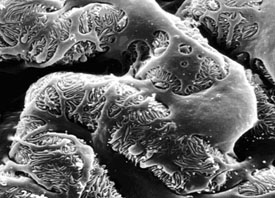
Scanning electron micrograph of a podocyte. |
A glomerulus is essentially a complex capillary tuft comprised of a number of unique cell types. One of these varieties - podocytes, are epithelial cells exhibiting an exquisite morphology consisting of interdigitating foot processes extending from primary processes and the cell body - in fact, they look a lot like octopuses. The podocytes form the final filtration barrier that restricts the passage of albumin, while allowing water and small solutes to enter the nephron. Over the past few years, the renal research community has come to realize that these podocytes are primary targets of the majority of glomerular diseases - including diabetic nephropathy, HIV nephropathy, focal glomerulosclerosis and many others.
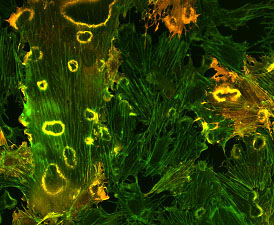
Cells expressing FSGS-associated α-actinin-4 form fewer ruffles. Additionally the emergence of these ruffles is delayed. |
Dr. Kennedy's lab is focused on the biology of the podocyte in the context of kidney disease. His group has engineered several transgenic mouse models of kidney disease. One model allows either overexpression or deletion of the prostaglandin EP4 receptor in podocytes, and is used in renal ablation models of glomerulosclerosis. The other is a model of focal glomerulosclerosis (FSGS, or glomerular scarring), an inherited form of FSGS which arises from a mutation in the cytoskeletal crosslinking protein, α-actinin-4. These mice proved that this form of FSGS is initiated in podocytes, and provided clues about how the disease develops.
Biographical Sketch
Dr. Chris Kennedy received his Ph.D. from the University of Ottawa in 1996 under the supervision of Drs. Pierre Proulx and Richard Hébert. He carried out his postdoctoral training in the Division of Nephrology at Vanderbilt University, under the supervision of Dr. Richard Breyer, from 1996 to June 2000. He arrived in Ottawa in July 2000 and is currently a Research Scientist within the Kidney Research Centre at the Ottawa Health Research Institute. He is married to Pam Kennedy, a piano teacher, and has two energetic children (Cassidy, 2 years old, and CJ, 9 months old).
For more information, visit Dr. Kennedy's website or the OHRI's Kidney Research Centre.
| |

