Flow Cytometry and Cell Sorting
Instrumentation: SONY MA900
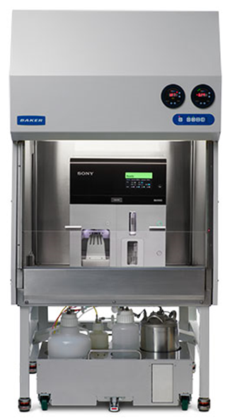
Sony MA900 Cell Sorting instrument
Location: The Ottawa Hospital Cancer Centre, 3rd floor, South Wing, room 3340E, 501 Smyth Road
The SONY MA900 is a user-operated cell sorting instrument capable of detecting up to 12 different fluorescent markers, plus forward and side scatter parameters, and the ability to sort into different size tubes and plates.
Optics specifications:

The SONY MA900 is equipped with 4 excitation lasers (405 nm, 488 nm, 561 nm, 638 nm) in a dual axis optical system, (colinear system between 488 / 561 nm lasers and 405 / 638 nm lasers.
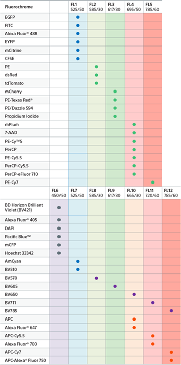
The instrument is capable of detecting up to 12 different fluorescent parameters (5 parameters in 488 / 561 nm lasers and 7 parameters in 405 / 638 nm lasers) plus forward and side scatter parameters.
Fluidics and sorting specifications:
The instrument uses an auto loading chamber where you can load your samples in different tube sizes, ranging from 0.5 mL to 15 mL. The loading chamber is also temperature controlled and has an agitation unit to maintain the cells in suspension during the sort.
The sorting chamber also allows for different sizes of tubes, from 0,5 mL to 50 mL, and it is capable of doing from 2-way to 4-way sorts. The sorting chamber has outstanding set up for different size plates, from 6- to 384-well plates.
The main feature of the SONY MA900 instrument is the microfluidics chip-based technology, which helps users to easily install and set up the chip/nozzle system into the instrument and automatically calibrate it. The facility has 3 different size nozzle chips which can be used depending on the type of cells the users will be sorting:
- 70 um nozzle
- 100 um nozzle
- 130 um nozzle
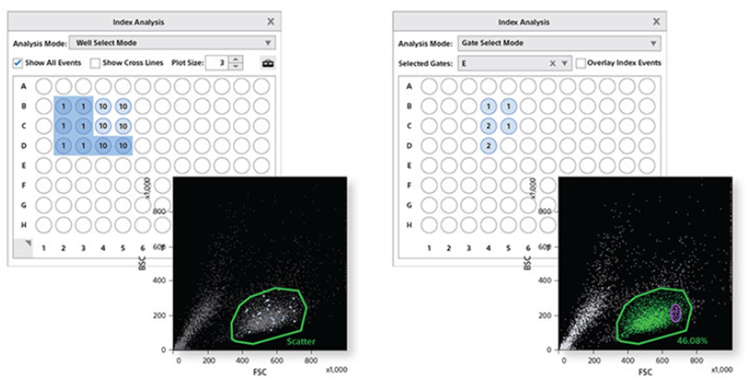
Index Sorting feature:
Index sorting software records the X and Y coordinates of each event sorted into a multi-well device. This very precise and easy to use software brings powerful capabilities to research, enabling you to track the scatter and fluorescence intensity of individual cells sorted in each well.
These index sort options allow you to perform meta-analysis of data for several applications. For example, clonal variability can be studied based on the expression levels of the fluorescent protein or surface markers. Also, researchers can integrate phenotypic data with mRNA expression analysis of the sorted cells.

Fluorochrome detector panel configuration:

